Anti-Candidal Activities of Leaf Extracts of Sansevieria aethiopica (Thunb): A Medicinal Plant Use in the Treatment of Oral Candidiasis in Eastern Cape of South Africa
Oluwole Moses David and Anthony Jide Afolayan
Oluwole Moses David1* and Anthony Jide Afolayan2
1Department of Microbiology, Ekiti State University, Ado-Ekiti Nigeria
2Medicinal Plants and Economic Development Research Centre, Department of Botany, University of Fort Hare, Alice 5700, South Africa
- *Corresponding Author:
- Oluwole Moses David
Department of Microbiology, Ekiti State University, Ado-Ekiti, Nigeria
Tel: +2348030883124
E-mail: davidoluwole5@gmail.com
Received date: May 02, 2016; Accepted date: December 23, 2016; Published date: January 05, 2017
Citation: David OM, JideAfolayan A. Anti-candidal Activities of Leaf Extracts of Sansevieria aethiopica (Thunb): A Medicinal Plant use in the Treatment of Oral Candidiasis in Eastern Cape of South Africa. J Pharm Microbiol. 2017, 3:1.
Abstract
Candida albicans infections are on the increase in the recent time and its resistance to most fungicides has been documented. We studied the effect of extracts of Sansevieria aethiopica (Thunb) on C. albicans ATCC 10231 and their possible mechanisms of actions were proposed. The minimum inhibitory concentrations (MICs) and minimum fungicidal concentrations (MFCs) of the extracts were determined using macrobroth dilution method while the structural changes in the fungus after treatment with the extract was determined by electron microscope. Standard methods were used to determine the effects of the extracts on the proton pumping, internal pH and ergosterol synthesis. The MIC of the extracts ranged from 1.5625 to 3.125 mg/ml while extracts-treated cells showed alterations in the morphology; wrinkled surfaces, shrinkages, tears and holes. Proton pumping activity was lower in the treatment group compare to the control while the internal pH of test fungus ranged between 5.40 and 6.03. We observed a decreased in ergosterol content in the candidal cells treated with the plant extracts. At ½MIC of acetone, methanol and ethanol extracts of the plants the amount of 24(28)-dihydroergosterol to ergosterol were 0.0972/0.5128, 0.0939/0.3571 and 0.1032/0.3702 g/dry weight respectively. The extracts were able to inhibit the growth, affect the intracellular pH (by extension the membrane integrity) and interfere with the sterol metabolism in C. albicans ATCC 10231.
Keywords
Candida albicans, Sansevieria aethiopica, ergosterol, proton pumping, intracellular pH, fungicide
Introduction
Candida albicans is a fungus usually reproduced by budding it is a normal flora of human gastrointestinal and vaginal tracts [1]. The occurrence of candidiasis is lower than the bacterial infections; however Candida albicans infections ranked fourth among nosocomial bloodstream infections leading to death [2-4]. Candida spp. are frequently colonizes mucous membrane of human where they act as opportunistic fungal pathogens which may develop to candidaemia [5,6]. The predisposing factors have been identified to include: high diabetes, pregnancy, usage oral contraceptives and antibiotics [7,8]. Candidal infections have increased significantly in the recent time with mortality rate of 40% [6-9]. Candida infection has been reported to be the one of the commonest causes of vaginitis among middle age women [10,11].
Medicinal plants are used for the treatment of contagious and physiological diseases globally. The applications of medicinal plants are prominent among rural dwellers in poor-resource nations [12,13]. The global report showed an increase in the resistance of disease causing microorganisms to different orthodox medicines [14-16]. This in no doubt called for alternative and effective therapy.
The use of medicinal plants has gained a wide recorgnition as a result of its safety, low cost and effectiveness [17,18]. This now shifted the focus to medicinal plants for the treatment of infections. These attributes make medicinal phytomedicine to be preferred to the conventional chemotherapeutics agents [17-20].
Sansevieria aethiopica (Thunb) is a perennial shrub with tough and erected leaves [21] used for the treatment of oral, ear and other fungal infections [22-23]. Despite its popular uses of S. aethiopica its mechanisms of actions have not been reported hence the aim of this study. We proposed the possible mechanisms of actions of S. aethiopica against C. albicans in this study.
Materials and Methods
Source and extraction of plant sample
Fresh leaves of S. aethiopica were collected from a single tuft in February, 2012, in Alice Township, Nkokobe Municipality of Eastern Cape of South Africa. At the Giffen Herbarium of the Department of Botany, University of Fort Hare, Alice, South Africa, the plant was authenticated by Prof. D. Grierson and the voucher (DavMed 2012/2) was submitted to the same Herbarium. The plant was dried in the oven at 40 °C and pulverized to fine powder and a 50 g of ground plant sample was soaked in 500 ml of each of the solvents for 12 h on Stuart Scientific Orbital Shaker (Manchester, UK). The sample was then suction-filtered through Whatman number 1 filter paper and washed with another 200 ml solvent. The filtrate was concentrated with Laborata 4000-efficient (Heldoph, Germany). Each of the dried extracts was dissolved in water + 2% dimethyl sulfoxide (DMSO). The reconstituted extracts were filter by 0.45 μl pore size membrane filter for sterility.
Source of the organism
Candida albicans ATCC 10231 was collected from the Department of Biochemistry and Microbiology, University of Fort Hare, Alice, South Africa. The organism was grown in Potato Dextrose Broth (PDB) for 18 h and was standardized to 0.5 McFarland scale (≈1.0 x 10⁷ cfu/ml) and diluted with sterile PDB to achieve the final concentration of 1.0 x 10⁶ cfu/ml.
Determination of minimum inhibitory concentration (MIC)
Macrobroth dilution method of Clinical and Laboratory Standards Institute (CLSI) [24] was used to determine the in vitro antifungal activity of the extracts of S. aethiopica. Extract of the plant sterilized by membrane filtered was incorporated into Potato Dextraose Broth in test tubes to give a final concentrations ranging from 0.078125 to 10.00 mg/ml. The tubes were inoculated with 100 μl of standardized inoculum of C. anlbicans ATCC 10231. A tube Amphotericin B and with only broth were used as a positive and negative controls respectively. The tubes were incubated aerobically at 37 °C for 24 h. The first tube in the series with no sign of visible growth was taken as the MIC.
Determination of minimum fungicidal concentration (MFC)
A loopful of culture from the first three broth tubes that showed no growth in the MIC tubes were inoculated on sterile PDA plates and observed for growth after incubation at 37 °C for 24 h. The MFC was taking as the least concentration of the extracts that showed no growth. MIC index (MICI) was calculated as the ratio of MFC and MIC. The result was interpreted as follow: MFC/MIC ≤2.0 was considered bactericidal, if ÃÆââ¬Â¹ÃâÃâ2 but ÃÆââ¬Â¹Ãâââ¬Å¡16 it was considered bacteriostatic and lastly if the ratio is ≥16.0, the extract was considered ineffective as reported by Shanmughapriya et al. [25].
In situ electron microscopy
The effects of the extracts of S. aethiopica on the ultra-structure of the C. albicans ATCC 10231 was determined by the method of Kamilla et al. [26] with minor modifications. Standardized inoculum of C. albicans ATCC 10231 (with optical density of 0.1 at the wavelength of 650nm) was seeded on a sterile plate of Potato Dextrose Agar and incubated for 2 h at 37 °C. The plant extracts at different minimum fungicidal concentrations was applied on the culture and the plate was further incubated for another 6 h at 37 °C. Five mm sterile borer was used to remove plugs from the culture and each of the plugs was placed on a double-stick adhesive tab on a stub. The sample was vapour fixed with 2% osmium tetroxide for 1 h, dried with liquid nitrogen and later transferred to freeze dryer (VisTis Benchtop K) for 5 h. The dried samples were gold coated before viewing with scanning electron microscope (JEOL JSM-6390LV, Japan).
Proton pumping activity
The method of Shreaz et al [27]. was used to determine the effect of the extracts on the proton pumping in Candida albicans ATCC 10231. The test fungus was culture in Yeast Nitrogen Broth supplemented with 20 % glucose and grown for 18 h at 30 °C with periodic shaking. After incubation the cells were harvested by centrifuging at 3500 × g for 10 min at 4 °C and washed twice with sterile distilled water. Harvested cells (0.1 g) were re-suspended in 5 ml solution containing 0.1 M KCl and 0.1 mM CaCl2. The ¼ MIC and ½ MIC of extracts of S. aethiopica were added separately into the suspension after which the pH was adjusted to 7.0 using 0.01 M HCl/NaOH. Dimethyl sulfoxide (2%) and Amphotericin B (6 μg/ml) were used as negative and positive control respectively in the assay. The pH of the suspension was adjusted to 7.0 using 0.01 M HCl/NaOH. Test extracts were added to achieve the desired concentrations (¼ MIC and ½ MIC) in 5 ml solution. A 100 ml of glucose was added to the suspension to achieve a final concentration of 5 mM and the pH of the suspension was monitored, over a period of 60 min, using Crison Basic 20 pH meter (Barcelona, Spain).
Measurement of intracellular pH (pHi)
Modified method of Bhatia et al [28]. was used to determine the intracellular pH of the test fungus. As described above, C. albicans ATCC 10231 cells were grown, harvested and washed twice with sterile distilled water as described earlier and 0.1 g of the cells was suspended in 5 ml 50 mM KCl. The extract of S. aethiopica was added at sub MICs in each case and Amphotericin B was also added to the control group. The pH of the medium was adjusted to 7.0 using Crison Basic 20 pH meter (Barcelona, Spain) and incubated at 37 °C for 30 min with constant shaking. The pH of the medium was monitor and the point of no further change in the pH was taking as the internal pH.
Determination of ergosterol content in the plasma membrane
Total intracellular sterols were extracted as reported by Arthington- Skaggs et al [29]. with slight modification. Standardized inoculum of C. albicans ATCC 10231 was plated in Potato Dextrose Broth (PDB) medium containing sub-MICs of extracts of S. aethiopica except, the control, and incubated for 24 h at 37 °C. The cells were harvested by centrifugation at 2,700 rpm for 5 min washed twice with distilled water and cell pallets weighed. A 5 ml of 25% alcoholic potassium hydroxide (25 g of KOH and 36 ml of sterile distilled water, brought to 100 ml with 100% ethanol) solution was added to each sample and vortex mixed for 2 min and incubated in an 80 °C water bath for 1 h. After incubation, tubes were allowed to cool to room temperature and 2 ml of sterile distilled water and 5 ml of n-heptane, followed by vigorous vortex mixing for 3 min. The samples were stored at -20 °C for 24 h. The n-heptane layer was scan spectrophotometrically by UV-VIS 3000PC over a wavelength range of 220 and 300 nm. Ergosterol and late sterol intermediate 24(28) dehydroergosterol (DHE) in the extracted sample were quantified from the result as followed Ergosterol content was calculated as a percentage of the wet weight of the cell by the following equations:
% Ergosterol + %24(28) DHE = (A282/290)/pellet weight
%24(28) DHE = (A230/518)/pellet weight
% Ergosterol = % Ergosterol + %24(28) DHE - %24(28) DHE
Where 290 and 518 are E value (in percentage per cm) determined for crystalline ergosterol and 24(28)dehydroergosterol respectively.
Statistical Analyses
All the experiments were conducted in triplicates and repeated twice. Data are shown as mean ± standard deviation (SD) for quantitative variables and as absolute qualitative variables. Analyses were conducted by Dunnett Multiple Comparisons Test to compare both treatment groups with the control using SPSS version 17.0 (SPSS, Chicago, IL, USA).
Results and Discussion
The search for new antifungal drugs is very necessary in the face of current resistance of pathogens to the present chemotherapy [30]. The MIC of the extracts ranged between 1.5625 and 3.125 mg/ml. Acetone extract had the highest MIC of 3.125 mg/ ml and both Acetone and ethanolic extracts had MFC of 6.250 mg/ml as shown in Table 1. Acetone and methanolic extracts had fungicidal effects on the test yeast. Plant extracts or plantderived compounds have been very effective against Candida spp. This results support the reports of other researchers that plant extracts are very effective in the treatment of candidiasis [16,18,20].
| Extracts | MIC | MFC | MFC/MIC | Interpretation |
|---|---|---|---|---|
| Acetone | 3.125 | 6.250 | 2 | Fungicidal |
| Ethanol | 1.5625 | 6.250 | 4 | Fungistatic |
| Methanol | 1.5625 | 1.5625 | 1 | Fungicidal |
| Amphotericin B (mg/ml) | 0.0080 | 0.0160 | 2 | Fungicidal |
Table 1: Antifungal activity of extracts of S. aethiopica (mg/ml) on C. albicans ATCC 10231.
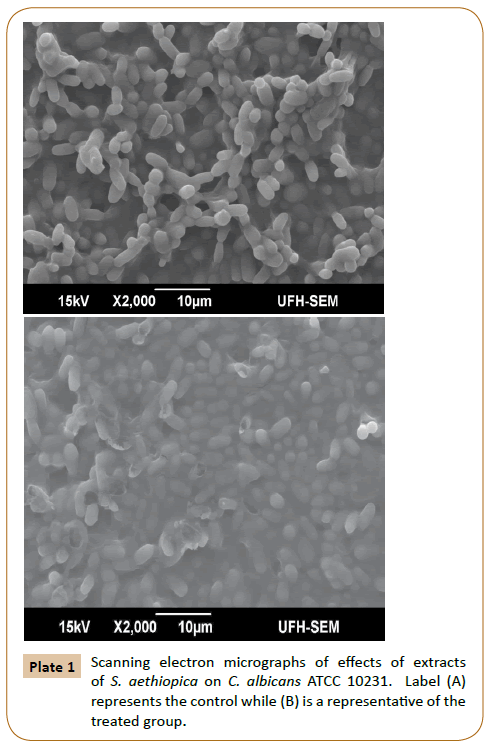
The cells in the control plate are with a normal, round and smooth-surfaced while those in the control in the treated plate showed alterations in the cell morphology (Plate 1). In the plate B there were burst cells that have released out the cytoplasmic contents. Treated cells showed changes in their morphology, a number of damaged cells features were shown. Wrinkled surfaces, shrinkages, tears and holes in the cells were observed in the treated cells. The effects of the extracts of S. aethiopica with cell membrane integrity as evidenced by shrinkage of cell surface and lysis of sessile cells indicate their effectiveness in the eradication of C. albicans. This is similar to reports of other researchers that have indicated the compounds from plants exert disruption of cell membrane of the yeast cells [31,32]. The changes in membrane morphology of Candida albicans ATCC 10231 treated with extracts of S. aethiopica we observed is similar to the findings of Pan et al [33]. who reported extracellular material and membrane perturbations as a result of membranes destruction in C. albicans.
Glucose-dependent medium acidification provides a relative measure of proton pumping by the plasma membrane. The effect of extracts of S. aethiopica on glucose-dependent proton pumping by C. albicans ATCC 10231 was evaluated. Compared with the control, there was a significant difference in the data obtained in the treatment groups: ethanolic and methanolic extracts of S. aethiopica at ½ MIC and ¼ MIC as shown in Table 2. Glucose dependent proton pumping showed a dose-and timedependent inhibition of proton pumping. The proton-pumping ability of fungi is crucial for the regulation of the internal pH of a fungal cell. When fungal cells depleted of their carbon sources are exposed to glucose, the sugar is rapidly taken up by the cells by the proton motive force generated by the proton gradient due to the pumping out of intracellular protons [27,34].
| Extracts | Conc. | Time in minutes | ||||||
|---|---|---|---|---|---|---|---|---|
| 0 | 10 | 20 | 30 | 40 | 50 | 60 | ||
| Ethanolic | ½ MICa,e,f | 7.00 | 6.25 ± 0.13 | 6.21 ± 0.23 | 6.17 ± 0.19 | 6.02 ± 0 ± 0.73 | 5.98 ± 0.67 | 5.51 ± 0.49 |
| ¼ MIC | 7.00 | 6.23 ± 0.46 | 6.11 ± 0.45 | 6.10 ± 0.82 | 5.82 ± 0.93 | 5.77 ± 0.43 | 5.62 ± 0.92 | |
| Methanol | ½ MICb,f | 7.00 | 6.34 ± 0.34 | 5.99 ± 0.64 | 5.73 ± 0.73 | 5.44 ± 0.46 | 5.32 ± 0.82 | 4.70 ± 0.84 |
| ¼ MICc,f | 7.00 | 6.33 ± 0.47 | 5.83 ± 0.69 | 5.65 ± 0.46 | 5.4 ± 0.48 | 5.26 ± 0.73 | 4.89 ± 0.45 | |
| Acetone | ½ MICd,f | 7.00 | 6.32 ± 0.55 | 5.74 ± 0.93 | 5.48 ± 0.37 | 5.21 ± 0.29 | 4.93 ± 0.48 | 4.39 ± 0.45 |
| ¼ MICe,a,f | 7.00 | 6.12 ± 0.11 | 5.53 ± 0.28 | 5.37 ± 0.12 | 5.02 ± 0.46 | 4.32 ± 0.25 | 4.15 ± 0.45 | |
| Controlf,a,b,c,d | 7.00 | 5.82 ± 0.92 | 5.25 ± 0.72 | 5.00 ± 0.84 | 4.73 ± 0.99 | 4.23 ± 0.37 | 4.01 ± 0.57 | |
Table 2 Effect of extracts of S. aethiopica on proton pumping of C. albicans ATCC 10231.
The effect of extracts on the proton-pumping ability of C. albicans was determined by glucose-induced acidification of external medium. The extracts inhibited the glucose-induced acidification of the external medium by C. albicans in a concentrationdependent manner. As shown in Table 2, at ½MIC the rate of inhibition of glucose-induced acidification of the external medium by C. albicans, measured in term of the pH of the medium, within 30 min of the experiment ranged between 6.25-5.51, 6.34-4.70 and 6.32-4.39 for ethanolic, methanolic and acetone extracts respectively. Paired Samples Statistics (Paired T-test) at probability level of 0.05 showed a significance difference between the control and the three test groups treated at the ½MIC. The activity of the lower concentration ¼MIC was expectedly lower that the ½MIC in all the treatment groups.
Regulation of intracellular pH is a fundamental to the growth of Candida and activation of plasma membrane ATPase as it is involved in maintenance of pHi [35,36]. From the Table 3, the internal pH of test fungus ranged between 5.87 and 6.03 for the cells treated with ¼MIC. The value was lower in the cells treated with ½MIC than those treated with ¼MIC of the plant extracts. For the cells treated with ½MIC the highest pHi was recorded in ethanolic extract treated cells while the least was observed in the methanolic extracts. The decrease in pHi was more in cells exposed to the extracts compared with the control. Prolonged intracellular acidification has been reported to have deleterious effect on cell viability [37,38]. This also affects the efflux of potassium ions [39] and cellular ion homeostasis [37,40].
| Extracts | Concentrations | ||
|---|---|---|---|
| ¼ MIC | ½ MIC | ||
| Acetone | Mean | 5.91 | 5.40 |
| SEM | 0.34 | 1.24 | |
| Ethanol | Mean | 6.03 | 5.52 |
| SEM | 1.01 | 0.72 | |
| Methanolic | Mean | 5.87 | 5.24 |
| SEM | 0.32 | 0.02 | |
| Control (Amphotericin B) | Mean | 7.38 | |
| SEM | 0.34 | ||
Table 3: Intracellular pH (pHi) in presence of the extracts of S. aethiopicain C. albicans ATCC 10231.
Sterol quantification has been reported by Arthington-Skaggs et al [30]. To be more predictive of in vivo outcome than the broth microdilution procedure. We noticed a decrease in the level of the ergosterol and 24(28)DHE in the treated cell compared to the control. The amounts of the sterols were least in the cell treated with ½MIC of the extract of S. aethiopica. This showed a concentration dependent effect of the plant extracts. At the ½MIC of acetone, methanol and ethanol the amount of 24(28)DHE/ergosterol were 0.0972/0.5128, 0.0939/0.3571 and 0.1032/0.3702 respectively (Table 4). This finding has been corroborated by other researchers [41-43]. Ergosterol is a significant component of Candida spp. and its content is affected by chemical and physical factors [44].
| Extract | Pallet weight (g) | Percentage of the wet weight | |||
|---|---|---|---|---|---|
| Ergosterol + 24(28)DHE | 24(28)DHE | Ergosterol | |||
| Acetone | ½MICa | 1.23 ± 0.13 | 0.4619 ± 0.0302 | 0.0798 ± 0.0016 | 0.3822 ± 0.0240 |
| ¼MIC | 2.49 ± 0.36 | 0.6100 ± 0.0023 | 0.0972 ± 0.0035 | 0.5128 ± 0.0722 | |
| Methanol | ½MICb | 1.28 ± 0.11 | 0.2883 ± 0.0104 | 0.0843 ± 0.0204 | 0.2040 ± 0.0492 |
| ¼MICc | 2.01 ± 0.09 | 0.4510 ± 0.0033 | 0.0939 ± 0.0310 | 0.3571 ± 0.0240 | |
| Ethanol | ½MICd | 1.39 ± 0.14 | 0.2655 ± 0.0308 | 0.0776 ± 0.0100 | 0.1879 ± 0.0085 |
| ¼MIC | 2.87 ± 0.40 | 0.4734 ± 0.0200 | 0.1032 ± 0.0378 | 0.3702 ± 0.0449 | |
| Controla,b,c,d | 4.81 ± 1.73 | 1.4786 ± 0.0382 | 0.8203 ± 0.2391 | 0.6583 ± 0.0231 | |
Table 4: Inhibition of ergosterol biosynthesis in C. albicans ATCC 10231.
We observed a decreased in ergosterol content Candida cells treated with the plant extracts. The difference in the activity of the extract may be due to their difference in terms of interactions with cholesterol as reported by Henriksen et al [45]. This is as a result of interference with the sterol metabolism which is the primary cellular process. This shows that the extracts target the cell membranes of Candida albicans. In conclusion, the extracts of S. aethiopica have been able to inhibit the growth of C. albicans ATCC 10231 and also we observed that the extracts were able to affect the intracellular pH and interfere with the sterol metabolism. This plant has multiple actions on the cell wall of the test fungus.
Acknowledgements
The authors wish to acknowledge the financial supports of the National Research Foundation, South Africa and the University of Fort Hare, Alice 5700, Eastern Cape, South Africa.
References
- Doyle TC, Nawotka KA, Kawahara CB, Francis KP, Contag PR (2006) Visualizing fungal infections in living mice using bioluminescent pathogenic Candida albicans strains transformed with the firefly luciferase gene. Microb Pathog 40: 82-90.
- Beck-Sague C, Jarvis WR (1993) Secular trends in the epidemiology of nosocomial fungal infections in the United States, 1980–1990. National nosocomial infections surveillance system. J Infect Dis 167: 1247-1251.
- Edmond MB, Wallace SE, McClish DK, Pfaller MA, Jones RN, et al. (1999) Nosocomial bloodstream infections in United States hospitals: a three-year analysis. Clin Infect Dis 29: 239-244.
- Pfaller MA, Diekema DJ (2007) Epidemiology of invasive candidiasis: a persistent public health problem. Clin Microbiol Rev 20: 133-163.
- Lipman J, Saadia R (1997) fungal infections in critically ill patients. Br Med J 315: 266-267.
- Pfaller M, Wenzel R (1992) Impact of the changing epidemiology of fungal infections in the 1990s. Eur J Clin MicrobInfect Dis 11: 287-291.
- Leigh DA, Joy GE, Tait S, Harris K, Walsh B (1989) Treatment of acute uncomplicated urinary tract infections with single daily doses of cefuroxime axetil. J Antimicrob Chemother 23: 267-73.
- Ascioglu S, Rex JH, Pauw B, Benett JE, Bille J, et al. (2002) Defining opportunistic invasive fungal infections in immunocompromised patients with cancer and hematopoietic stem cell transplants: an international consensus. Clin Infect Dis 34: 7-14.
- Klevay MJ, Horn DL, Neofytos D,Pfaller MA, Diekema MJ (2009) Initial treatment and outcome of Candida glabrata versus Candida albicans bloodstream infection. Diagn Microbiol Infect Dis 64: 152-157.
- Harriott MM, Lilly EA, Rodriguez TE, Fidel PL, Noverr MC (2010) Candida albicans forms biofilms on the vaginal Mucosa. Microbiology 156: 3635-3644.
- Lopez-Medrano F, Fernandez-Ruiz M, Origuen J (2012) Clinical significance of Candida colonization of intravascular catheters in the absence of documented candidemia. Diagn Microbiol Infect Dis 73: 157-161.
- Duraipandiyan V, Ignacimuthu S (2011) Antifungal activity of traditional medicinal plants from Tamil Nadu, India. Asian Pacific J Trop Biomed S204-S215.
- Wintola OA, Afolayan AJ (2015) an inventory of indigenous plants used as anthelmintics in Amathole District Municipality of the Eastern Cape Province, South Africa. Afr J Tradit Complement Altern Med 12: 112-121.
- Govindachari TR, Suresh G, Gopalakrishnan G, Balaganesan B, Masilamani S (1998) Identification of antifungal compounds from the seed oil of Azadirachta indica. Phytoparasitica 26: 109-116.
- Srinivasan D, Nathan S, Suresh T, Lakshmana PP (2010) Antimicrobial activity of certain Indian medicinal plants used in folkloric medicine. J Ethnopharmacol 74: 217- 220.
- Tharkar PR, Tatiya AU, Shinde PR, Surana SJ, Patil UK (2010) Antifungal activity of GlycyrrhizaglabraLinn. andEmblicaofficinalisGaertn. byeirectbioautography method. Int J Phar Tech Res 2: 1547-1549.
- Parekh J, Chanda S (2008) In vitro antifungal activity of methanol extracts of some Indian medicinal plants against pathogenic yeast and moulds. AfrJ Biotechnol 7: 4349-4353.
- Patel M, Coogan MM (2008) Antifungal activity of the plant Dodonaeaviscosavar. Angustifolia on Candida albicans from HIV-infected patients. J Ethnopharmacol 118: 173-176.
- Gupta SK, Banerjee AB (2008) Screening of selected West Bengal plants for antifungal activity. Econ Bot 26: 255-259.
- Suresh M, Rath PK, Panneerselvam A, Dhanasekaran D, Thajuddin N (2010) Antifungal activity of selected Indian medicinal plant salts. J Global PharmaTechnol 2: 71-74.
- Van Wyk BV, Gericke N, Van Oudtshoorn B (2000). Medicinal Plants of South Africa. Briza Publications Pretoria South Africa.
- Hutchings A, Scott AH, Lewis G, Cunningham A (1996) Zulu Medicinal Plants. University of Natal Press, Pietermaritzburg.
- Newton LE (2001) Sansevieria. In: Succulents Encyclopedia. Volume 1 monocots plants (monocots), Eugen Ulmer, Stuttgart, p272.
- Clinical and Laboratory Standards Institute (CLSI) (2005) Performance standards for antimicrobial susceptibility testing. 15th informational supplement. Wayne, PA: Clinical and Laboratory Standards Institute; M100-S15.
- Shanmughapriya SA, Manilal A, Sujith S, Selvin J, Kiran GS, et al. (2008) Antimicrobial activity of seaweeds extracts against multiresistant pathogens. Ann Microbiol 58: 535-541.
- Kamilla L, Mansor SM, Ramanathan S, Sasidharan S (2009) Effects of Clitoria ternatea leaf extract on growth and morphogenesis of Aspergillus niger. Microsc Microanal 15: 366-372.
- Shreaz S, Bhatia R, Khan N, Ahmad SI, Muralidhar S, et al. (2011) Interesting anti-candidal effects of anisic aldehydes on growth and proton-pumping-ATPase-targeted activity. Microb Pathog 51: 277-284
- Bhatia R, Shreaz S, Khan N, Muralidhar S, Basir SF, et al. (2011) Proton pumping ATPase mediated fungicidal activity of two essential oil components. J Basic Microbiol 51: 1-9.
- Arthington-Skaggs BA, Warnock DW, Morrison CJ (2000) Quantitation of Candida albicans ergosterol content improves the correlation between in vitro antifungal susceptibility test results and in vivo outcome after fluconazole treatment in a murine model of invasive candidiasis. Antimicrob Agents Chemother 44: 2081-2085.
- Richardson MD (2005) Changing patterns and trends in systemic fungal infections. J Antimicrob Chemother 56: 5-11.
- Bennis S, Chami F, Chami N, Bouchikhi T, Remmal A (2004) Surface alteration of Saccharomyces cerevisiae induced by thymol and eugenol. Lett App lMicrobiol 38: 454-458.
- Tyagi AK, Malik A (2010) Liquid and vapour-phase antifungal activities of selected essential oils against Candida albicans: microscopic observations and chemical characterization of Cymbopogoncitratus. BMC Compliment Altern Med 10: 10-65.
- Pan C, Chen J, Lin T, Lin C (2009) In vitro activities of three synthetic peptides derived from epinecidin-1 and an anti-lipopolysaccharide factor against Propionibacterium acnes, Candida albicans, and Trichomonas vaginalis. Peptides 30: 1058-1068.
- Ben-Josef AM, Manavathu EK, Platt D (2000) Proton translocating ATPase mediated fungicidal activity of a novel complex carbohydrate: CAN- 296. Int J Antimicrob Agents 13: 287-95.
- Kauer S, Mishra P, Prasad R (1988) Dimorphism-associated changes in intracellular pH of Candida albicans BiochimBiophysActa 972: 277-282.
- Manzoor N, Amin M, Khan L (2002) Effect of phosphocreatine on H+ extrusion, pHi and dimorphism in Candida albicans. Int J ExpBiol 40: 785-790.
- Bagar T, Altenbach K, Read ND, Bencina M (2009) Live-cell imaging and measurement of intracellular pH in filamentous fungi using a genetically encoded ratiometric probe. Eukaryot Cell 8: 703-712.
- Plumridge A, Hesse SJ, Watson AJ, Lowe KC, Stratford M, et al. (2004) The weak acid preservative sorbic acid inhibits conidial germination and mycelial growth of Aspergillus niger through intracellular acidification. Appl EnvironMicrobiol 70: 3506-3511.
- Pena A, Calahorra M, Michel B, Ramirez J, Sanchez NS (2009) Effects of amiodarone on K+, internal pH and Ca2+ homeostasis in Saccharomyces cerevisiae. FEMS Yeast Res 9: 832-849.
- Bagar T, Bencina M (2012) Antiarrhythmic drug amiodarone displays antifungal activity, induces irregular calcium response and intracellular acidification of Aspergillus niger– Amiodarone targets calcium and pH homeostasis of A. niger. Fungal GenetBiol 49: 779-791.
- Arthington-Skaggs BA, Jradi H, Desai T, Morrison CJ (1999) Quantitation of ergosterol content: novel method for determination of fluconazole susceptibility of Candida albicans. J ClinMicrobiol 37: 3332-3337.
- Ghannoum M.A, Rice L.B (1999) Antifungal agents: mode of action, mechanisms of resistance, and correlation of these mechanisms with bacterial resistance. ClinMicrobiol Rev 2: 501-517.
- Dismukes WE (2000) Introduction to antifungal drugs. Clin Infect Dis30: 653-657
- Arnezeder CH, Koliander W, Hampel WA (1989) Rapid determination of ergosterol in yeast cells. Anal ChimActa 225: 129-136.
- Henriksen J, Rowat AC, Brief E, Hsueh YW, Thewalt JL, et al. (2006)Universal behavior of membranes with sterols. Biophys J 90: 1639-1649.
Open Access Journals
- Aquaculture & Veterinary Science
- Chemistry & Chemical Sciences
- Clinical Sciences
- Engineering
- General Science
- Genetics & Molecular Biology
- Health Care & Nursing
- Immunology & Microbiology
- Materials Science
- Mathematics & Physics
- Medical Sciences
- Neurology & Psychiatry
- Oncology & Cancer Science
- Pharmaceutical Sciences

